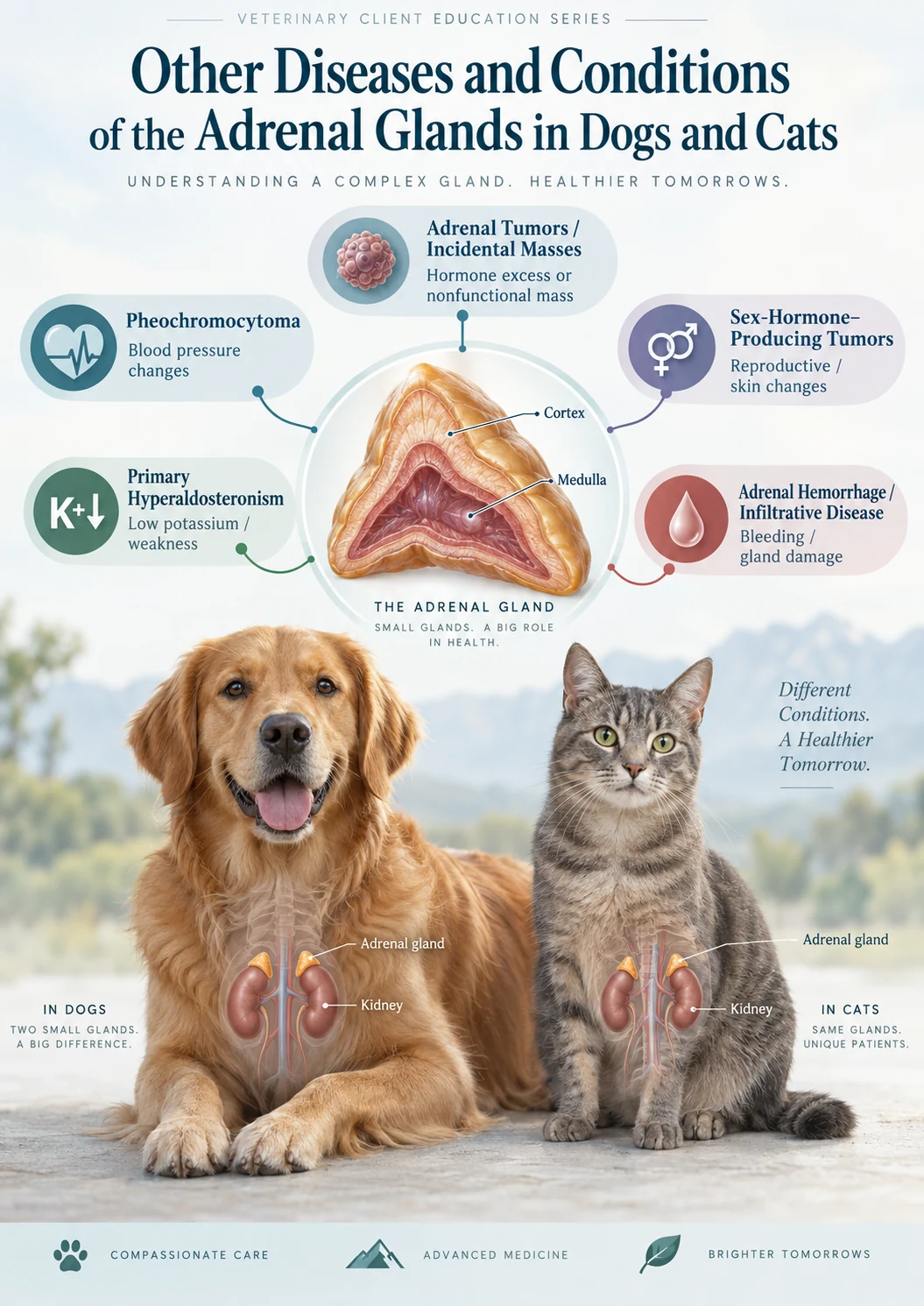
Other Diseases and Conditions of the Adrenal Glands in Dogs and Cats
Beyond Addison's Disease and Cushing's Disease
The adrenal glands are small organs, but they produce several hormones with powerful effects throughout the body. Addison's disease develops when adrenal hormone production becomes inadequate, while Cushing's disease involves excessive cortisol exposure. Those are the best-known adrenal disorders, but they are not the only diseases that can affect these glands.
Other adrenal conditions include tumors of the adrenal cortex or medulla, excessive production of aldosterone or sex hormones, adrenal masses that produce no hormone at all, hemorrhage within or around an adrenal gland, metastatic cancer involving the adrenal glands, and several uncommon inflammatory, infiltrative, or developmental disorders.
Some of these diseases cause obvious hormonal abnormalities. Others are discovered unexpectedly during an abdominal ultrasound or CT examination performed for an unrelated problem. Determining whether an adrenal abnormality is functionally active, locally invasive, malignant, or simply an incidental finding is often the most important part of the diagnostic process.
Understanding the Adrenal Gland
Each adrenal gland has two major regions with very different functions. The outer portion, the adrenal cortex, produces several steroid hormones. Cortisol influences metabolism, circulation, inflammation, immune function, and the response to stress. Aldosterone regulates sodium, potassium, water balance, and blood pressure. The adrenal cortex also produces smaller quantities of sex steroids and their precursors.
The inner portion, the adrenal medulla, contains specialized neuroendocrine cells that produce the catecholamines epinephrine and norepinephrine. These hormones participate in the “fight-or-flight” response by increasing heart rate, vascular tone, blood pressure, and availability of energy.
A disease arising from the adrenal cortex may therefore behave very differently from one developing in the adrenal medulla.
Adrenal Masses and Incidentalomas
Modern abdominal ultrasound and CT examinations identify adrenal nodules and masses much more frequently than they did in the past. An adrenal abnormality discovered during imaging performed for an unrelated problem is often called an adrenal incidentaloma.
Finding an adrenal mass does not automatically mean that the animal has adrenal cancer or even adrenal disease. Some adrenal nodules are benign and nonfunctional. Others produce excessive hormones. Some are primary adrenal tumors, while others represent cancer that has spread to the adrenal gland from another location.
Evaluating an Adrenal Mass
Evaluation commonly begins with the history, physical examination, blood pressure, CBC, chemistry profile, electrolytes, urinalysis, and assessment for recognizable endocrine patterns. The choice of hormone testing then depends on the clinical picture.
A dog showing signs of cortisol excess may be evaluated for Cushing's disease. A cat with hypertension and low potassium may require evaluation for excessive aldosterone production. A dog with episodic weakness, extreme blood-pressure changes, tachycardia, or collapse may require investigation for pheochromocytoma.
Ultrasound provides information about adrenal size, symmetry, internal architecture, and relationships with nearby structures. CT becomes particularly valuable when determining whether a tumor is invading major vessels or when surgery is being considered. Size alone cannot reliably determine whether an adrenal lesion is benign or malignant, although increasing size, irregular architecture, local invasion, vascular invasion, rapid growth, and evidence of metastatic disease increase concern.
Pheochromocytoma
A pheochromocytoma is a tumor arising from catecholamine-producing cells of the adrenal medulla. These tumors are uncommon in dogs and rare in cats, but they are clinically important because secretion of epinephrine and norepinephrine can produce dramatic cardiovascular effects. They also have a tendency to invade nearby vessels, particularly the caudal vena cava.
Pheochromocytomas usually occur in older dogs. Some are discovered incidentally before any recognizable symptoms develop.
Symptoms of Pheochromocytoma
Catecholamine secretion can be intermittent, so affected dogs may seem normal between episodes. Possible signs include weakness, restlessness, panting or rapid breathing, rapid heart rate, intermittent collapse, decreased appetite, weight loss, increased drinking and urination, abdominal discomfort, or unusual episodes of anxiety or agitation.
Hypertension is particularly important. Because catecholamine release may occur unpredictably, blood pressure can fluctuate considerably. Arrhythmias can occur, and severe cases may develop sudden cardiovascular instability or collapse.
Diagnosing Pheochromocytoma
An adrenal mass combined with unexplained hypertension or episodic cardiovascular signs raises suspicion, but ultrasound appearance alone cannot reliably distinguish a pheochromocytoma from an adrenal cortical tumor.
Blood pressure measurement is important. Biochemical testing can measure metabolites of epinephrine and norepinephrine known as metanephrines and normetanephrines. CT is particularly useful because it can define the mass and determine whether the tumor has entered the caudal vena cava or other major vessels. Thoracic imaging may also be recommended to look for metastatic disease.
Treatment
When the tumor is surgically resectable, adrenalectomy is the treatment of choice. These surgeries require careful planning because sudden catecholamine release during anesthesia or tumor manipulation can produce severe hypertension, arrhythmias, or cardiovascular instability. Vascular invasion can make surgery considerably more complicated.
Preoperative alpha-adrenergic blockade may be considered when a functional pheochromocytoma is suspected. Dogs that cannot undergo surgery may sometimes receive medical treatment aimed at controlling catecholamine effects and hypertension.
Primary Hyperaldosteronism
Primary hyperaldosteronism, also called primary aldosteronism or Conn's syndrome, develops when an adrenal gland produces excessive amounts of aldosterone independent of the body's normal regulatory system.
This disease deserves particular attention in cats and is probably underdiagnosed. Most recognized feline cases are associated with an aldosterone-producing adrenal cortical tumor, although bilateral adrenal hyperplasia can also occur. The disease is much less commonly recognized in dogs.
What Excess Aldosterone Does
Aldosterone instructs the kidneys to retain sodium while excreting potassium. Excessive aldosterone therefore commonly produces two major problems: potassium depletion and systemic hypertension.
Severe potassium deficiency interferes with normal muscle function. Affected cats may become weak, reluctant to walk or jump, develop a plantigrade stance, or become unable to hold the head normally. The characteristic downward position of the head and neck caused by muscular weakness is called cervical ventroflexion.
Hypertension can injure the eyes, brain, kidneys, and cardiovascular system. Some cats first present because of sudden blindness caused by hypertensive retinal hemorrhage or retinal detachment.
Diagnostic Evaluation
Primary hyperaldosteronism should be considered particularly in an older cat with unexplained or difficult-to-control hypertension, persistent hypokalemia, muscular weakness, cervical ventroflexion, or an adrenal mass.
A serum or plasma aldosterone concentration is measured. An aldosterone concentration that is inappropriately high in a hypokalemic patient is particularly concerning because low potassium would normally suppress aldosterone production. Measurement of plasma renin activity and calculation of an aldosterone-to-renin ratio can strengthen the diagnosis when this testing is available.
Ultrasound or CT is then used to examine both adrenal glands. The mere presence of an adrenal mass does not prove hyperaldosteronism because an adrenal tumor could instead be nonfunctional or could produce cortisol, catecholamines, or another hormone.
Treatment
For an appropriate cat with a localized unilateral aldosterone-producing adrenal tumor, adrenalectomy can provide definitive treatment. Before surgery, hypertension and potassium depletion should be stabilized.
Cats that are not surgical candidates can often be managed medically with a combination of spironolactone, potassium supplementation, and antihypertensive therapy such as amlodipine according to the individual patient's needs. Medical therapy does not remove the underlying tumor but can substantially improve potassium concentrations, blood pressure, muscle strength, and quality of life.
Adrenal Sex-Hormone-Producing Tumors
The adrenal cortex normally produces small amounts of androgenic, estrogenic, and progesterone-related steroids. Rare adrenal cortical tumors can produce excessive quantities of these hormones or their precursors.
Possible manifestations include symmetrical hair loss, thin or abnormal skin, increased pigmentation, mammary gland enlargement, vulvar enlargement, changes in external genitalia, altered sexual behavior, prostate enlargement, feminization of male animals, or masculinization of females.
Some dogs develop a Cushing-like appearance despite having conventional cortisol testing that does not demonstrate classic hypercortisolism.
Diagnosis
Diagnosis begins by recognizing the clinical pattern and identifying an adrenal abnormality. Specialized endocrine testing may measure progesterone, 17-hydroxyprogesterone, androstenedione, testosterone, estradiol, or other adrenal steroids when clinically appropriate.
Interpretation can be difficult because concentrations of several adrenal steroids may also increase in ordinary Cushing's disease, and an isolated abnormal laboratory result does not necessarily prove that the hormone is causing the clinical signs. For a clearly functional unilateral adrenal cortical tumor, adrenalectomy may offer definitive treatment when the tumor is surgically appropriate.
Nonfunctional Adrenal Cortical Adenomas and Carcinomas
Not every adrenal cortical tumor produces enough hormone to create a recognizable endocrine syndrome. A nonfunctional adrenal adenoma may remain clinically silent and be discovered during imaging for another condition.
A nonfunctional adrenocortical carcinoma can likewise produce few endocrine signs but can still become clinically important because of growth, invasion, hemorrhage, compression of nearby structures, or metastasis.
Adrenalectomy
Surgical removal may be recommended when a mass is functional, enlarging, invasive, strongly suspicious for malignancy, or otherwise likely to cause future problems. Adrenalectomy is specialized surgery because the glands lie immediately adjacent to major vessels and important abdominal structures.
Adrenal Hemorrhage
Bleeding can occur within an adrenal gland or in the tissues surrounding it. Potential causes include trauma, coagulation disorders, severe systemic illness, vascular injury, or hemorrhage associated with an adrenal tumor. In some cases, the cause is never definitively established.
A small focal hemorrhage may be discovered incidentally during imaging. More extensive hemorrhage can cause abdominal pain, weakness, anemia, low blood pressure, or collapse.
If both adrenal glands are severely damaged, loss of adrenal cortical function can eventually produce signs of hypoadrenocorticism. Bilateral adrenal hemorrhage is one uncommon cause of Addison's disease.
Diagnosis relies heavily on imaging together with CBC, coagulation testing, blood pressure, endocrine evaluation when indicated, and investigation for underlying neoplasia or systemic disease. Treatment is directed at cardiovascular stabilization, correction of the underlying cause, and management of adrenal hormone deficiency if it develops.
Inflammatory, Granulomatous, and Infiltrative Adrenal Disease
The adrenal glands can occasionally be damaged by severe inflammation or infiltration of abnormal cells. Granulomatous disease, fungal or other infectious disease, immune-mediated destruction, and infiltrative neoplasia can involve one or both glands. Cancer arising elsewhere in the body can also metastasize to the adrenal glands.
When enough adrenal cortex is destroyed bilaterally, the clinical result may ultimately be Addison's disease. The underlying process, however, is an infiltrative or destructive adrenal disorder rather than the more common presumed immune-mediated form of primary hypoadrenocorticism.
The clinical presentation depends on the underlying disease and may include weight loss, fever, lethargy, gastrointestinal signs, abnormalities elsewhere in the body, or endocrine failure. Imaging may reveal enlarged, irregular, nodular, mineralized, or otherwise abnormal adrenal glands, although imaging changes are not always specific.
Diagnosis may require investigation for systemic infection or cancer and, in selected patients, cytology, biopsy, surgery, or examination of another more safely accessible abnormal tissue.
Metastatic Cancer Involving the Adrenal Glands
The adrenal glands have a substantial blood supply, making them potential sites for metastatic tumor cells. Lymphoma and various solid tumors can infiltrate or metastasize to the adrenal glands.
An adrenal lesion in a patient with known cancer therefore must be interpreted differently from an isolated incidental nodule in an otherwise healthy animal. Bilateral extensive infiltration can theoretically reduce adrenal hormone production sufficiently to cause hypoadrenocorticism, although this is uncommon.
Evaluation generally includes staging of the primary cancer, assessment of both adrenal glands, endocrine testing when indicated, and determination of whether identifying the exact adrenal lesion would change treatment.
Other Rare Adrenal Medullary Tumors
Pheochromocytoma is the best-recognized tumor of the adrenal medulla, but several much rarer neuroendocrine tumors can arise from related tissues. These include neuroblastomas and ganglioneuromas. Related tumors known as paragangliomas can arise from similar neuroendocrine tissue outside the adrenal gland.
These tumors may produce problems through local growth, invasion, compression of nearby structures, or occasionally hormone production. Diagnosis generally depends on imaging and histopathology, sometimes supplemented by immunohistochemical testing.
When an Adrenal Abnormality Is Found on Ultrasound
An ultrasound report describing an adrenal nodule or mass understandably causes concern, but the next step should be deliberate rather than assuming the lesion is malignant.
The veterinarian considers the entire patient: whether the animal is drinking and urinating excessively, has persistent hypertension, has low potassium, experiences episodic weakness or collapse, has Cushingoid changes, has another known cancer, and whether the adrenal lesion is unilateral or bilateral.
The appearance of the opposite adrenal gland is also important. The lesion's relationship to the caudal vena cava and surrounding structures may affect the next diagnostic step and surgical planning.
A Practical Diagnostic Approach
Routine laboratory evaluation commonly includes CBC, serum chemistry, electrolytes, and urinalysis.
Blood pressure is particularly important because hypertension can accompany pheochromocytoma, primary hyperaldosteronism, and cortisol excess.
Hormonal testing is selected according to the suspected syndrome rather than ordering every possible adrenal test indiscriminately.
Ultrasound evaluates adrenal morphology and screens the remainder of the abdomen.
CT is particularly valuable for larger or potentially invasive masses and for surgical planning because it defines relationships with major vessels more accurately.
Thoracic imaging may be indicated when a malignant adrenal tumor is suspected.
Tests for metanephrines or normetanephrines may be used when pheochromocytoma is suspected. Aldosterone and renin-related testing may be appropriate when hyperaldosteronism is suspected. Cortisol testing is performed when the history suggests Cushing's or Addison's disease. Specialized steroid hormone panels may occasionally be used when a sex-hormone-producing adrenal tumor is suspected.
Why Adrenal Masses Require Careful Surgical Planning
Adrenal tumors can pose challenges disproportionate to their size. The adrenal glands lie close to the kidneys, renal vessels, aorta, caudal vena cava, and other important structures. Some tumors grow directly into the vena cava or form tumor-associated thrombi within it.
Pheochromocytomas add another risk because tumor manipulation may release catecholamines and abruptly alter blood pressure and heart rhythm. For these reasons, advanced imaging and referral to a surgeon experienced with adrenal disease are often appropriate when adrenalectomy is being considered.
When Adrenal Disease Becomes an Emergency
Many adrenal masses develop slowly, but some adrenal disorders can produce acute emergencies.
A pheochromocytoma can occasionally produce a sudden hypertensive or cardiovascular crisis. Severe hyperaldosteronism may cause profound potassium depletion or hypertensive injury. Adrenal hemorrhage can cause acute blood loss. Extensive bilateral adrenal destruction can result in an Addisonian crisis.
Prognosis
There is no single prognosis for “adrenal disease.” A small, stable, nonfunctional adrenal nodule may never cause clinical disease. A cat with a successfully removed unilateral aldosterone-producing tumor may have an excellent outcome. A dog with a surgically resectable pheochromocytoma can also have substantial long-term survival following successful adrenalectomy.
A malignant adrenal cortical tumor that has invaded major vessels or metastasized carries a more guarded prognosis.
Final Perspective
Adrenal disease extends well beyond Addison's and Cushing's disease. The adrenal cortex can produce excessive aldosterone or sex steroids, develop functional or nonfunctional adenomas and carcinomas, undergo hemorrhage, or be damaged by inflammatory, infectious, infiltrative, or metastatic disease. The adrenal medulla can develop pheochromocytoma and other uncommon neuroendocrine tumors.
At the same time, improved imaging means that veterinarians increasingly discover harmless or clinically insignificant adrenal nodules in older animals. The discovery of an adrenal abnormality is therefore the beginning of a diagnostic process rather than a diagnosis by itself.
The central questions are whether the lesion is functional, invasive, malignant, or clinically silent. Careful integration of clinical signs, blood pressure, laboratory testing, endocrine testing, ultrasound, CT, and—when appropriate—surgery or histopathology allows the most appropriate treatment to be selected while avoiding unnecessary intervention for incidental lesions.
Additional Resources
MSD Veterinary Manual — Overview of the Adrenal Glands
Comprehensive veterinary information concerning adrenal cortical and medullary physiology and disease.
MSD Veterinary Manual — Pheochromocytomas in Animals
Professional review of adrenal medullary tumors, clinical presentation, diagnosis, imaging, treatment, and prognosis.
MSD Veterinary Manual — Feline Primary Hyperaldosteronism
Professional review of aldosterone excess in cats, including hypertension, hypokalemia, diagnosis, imaging, surgery, and medical treatment.
American Animal Hospital Association — Selected Endocrinopathies Guidelines
Consensus recommendations covering major canine and feline endocrine diseases.
The Schwarzman Animal Medical Center — Internal Medicine
Specialty internal-medicine resources addressing adrenal and other endocrine disorders in dogs and cats.
Cornell University College of Veterinary Medicine
Veterinary endocrinology, diagnostic laboratory, internal medicine, and adrenal testing resources.
Today's Veterinary Practice
Peer-reviewed clinical reviews of endocrine disease, diagnostic imaging, and adrenal disorders.
Clinician's Brief
Practical veterinary reviews concerning adrenal masses, endocrine testing, adrenal surgery, and management of complicated endocrine disease.
This article provides general educational information for pet owners and is not a substitute for veterinary examination, diagnosis, or treatment. An adrenal mass discovered on ultrasound or CT does not by itself establish cancer or hormonal disease. Interpretation requires consideration of the animal's history, clinical signs, blood pressure, laboratory findings, endocrine testing, imaging characteristics, concurrent diseases, and changes over time. Animals with collapse, profound weakness, sudden blindness, severe abdominal pain, difficulty breathing, persistent vomiting, marked cardiovascular abnormalities, or other signs of critical illness require prompt veterinary evaluation.